Breadcrumb
- Home
- Faculty & Services
- Flow Cytometry
10-Color Flow Cytometry
Main navigation
- Faculty and Services
- Autopsy
- Bone and Soft Tissue Pathology
- Breast Pathology
- Cytogenetics
- Cytopathology
- Dermatopathology
- Electron Microscopy
- Flow Cytometry
- Gastrointestinal and Liver Pathology
- Head and Neck Pathology
- Hematopathology
- Molecular Pathology
- Muscular Dystrophy/Muscle Biopsy
- Neuropathology
- Renal Pathology
- Surgical Pathology
- Urologic Pathology
The immunopathology laboratory performs several categories of diagnostic testing including autoimmune disease serologies, flow cytometry, immunohistochemistry, and immunofluorescence microscopy.
Autoimmune disease serologies include screening for diseases such as systemic lupus erythematosus (SLE), Sjogren's syndrome, scleroderma, Wegner's granulomatosis and related vasculitides, autoimmune hepatitis, primary biliary cirrhosis, ulcerative colitis, celiac disease and others. Testing methodologies used include enzyme immunoassay direct and indirect immunofluorescence microscopy.
Flow cytometry is done on bone marrow, peripheral blood, fine needle aspirations, miscellaneous body fluids, lymph nodes and solid tumors. Flow cytometry has a wide range of uses from typing lymphomas and leukemias to quantitation of lymphocyte subsets in suspected immunodeficiency diseases.
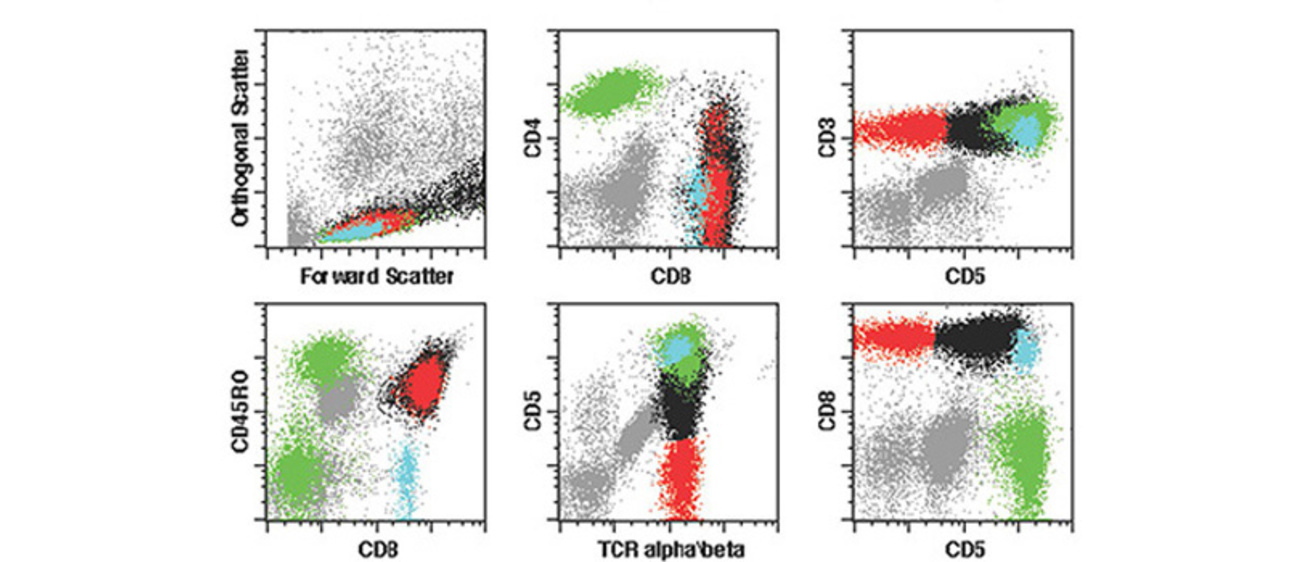
10-color flow cytometry employs both gating and cluster analysis strategies to maximize the sensitivity and accuracy of the testing.
Immunohistochemistry staining is done primarily on formalin-fixed, paraffin embedded tissues using automated staining equipment. A large number of antibodies are available to immunophenotype and identify the source of tumors and to detect certain infectious agents (particularly viruses) in tissue sections. In-situ hybridization studies are also performed to identify certain types on infections, particularly Epstein-Barr virus.
Immunofluorescence microscopy is done on frozen sections of the skin, mucosae and the kidney to diagnose autoimmune diseases.
How to Send Flow Cytometry Specimens
Hours: Specimens are accepted during our regular business hours.
Reports: Final result reports may be faxed or provided electronically.
Requisition form: Please print, fully complete, and send a Comprehensive Hematopathology Requisition with your specimens.
Consultants


Nitin Karandikar, MD, PhD
